Flexible Bronchoscopy

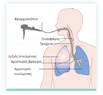
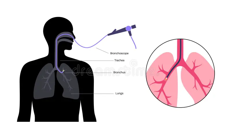
Flexible bronchoscopy is a medical procedure that involves the use of a flexible, thin tube with a light and a camera (bronchoscope) to examine the airways and lungs. The bronchoscope is typically inserted through the nose or mouth, allowing the healthcare provider to visualize the trachea, bronchi, and bronchioles in real-time. This procedure is commonly used for diagnostic purposes, such as obtaining tissue samples (biopsy) or performing bronchoalveolar lavage. Flexible bronchoscopy is a minimally invasive and versatile tool in pulmonology, aiding in the evaluation and diagnosis of various respiratory conditions, including infections, tumors, and lung diseases.
Why do I need a bronchoscopy?
- Diagnostic Evaluation:Bronchoscopy is used to investigate unexplained respiratory symptoms, such as persistent cough, wheezing, or hemoptysis (coughing up blood).
- Tissue Sampling (Biopsy):It allows for the collection of tissue samples from the airways, aiding in the diagnosis of lung infections, tumors, or inflammatory conditions.
- Visual Examination: Provides a direct visual inspection of the airways, enabling the identification of abnormalities, lesions, or obstructions.
- Treatment and Intervention: In certain cases, bronchoscopy can be used for therapeutic purposes, such as removing mucus plugs, placing stents, or treating airway strictures.
- Evaluation of Lung Diseases:Useful in assessing and diagnosing lung diseases, including pneumonia, interstitial lung disease, and bronchiectasis.
- Cancer Staging:Used in staging lung cancer to determine the extent of the disease within the airways and guide treatment decisions.
Exploring Alternatives to Bronchoscopy:
When bronchoscopy may not be the preferred approach, alternatives encompass chest imaging (CT scans), sputum cytology for cellular examination, pulmonary function tests, and the less invasive endobronchial ultrasound (EBUS) for visualizing the airways.
Preparing for Your Bronchoscopy:
Prior to the procedure, patients are typically instructed to adhere to a fasting period, follow specific pre-procedure guidelines, inform the healthcare team about medications, and arrange for post-procedure transportation due to potential sedation.
Navigating a Bronchoscopy Procedure:
During bronchoscopy, a flexible tube equipped with a camera is introduced through the nose or mouth to visually examine the airways. Biopsy samples may be collected, and therapeutic actions, such as addressing blockages, can be carried out.
Post-Bronchoscopy Procedure:
Following the bronchoscopy, patients undergo a short monitoring period in a recovery area. Temporary side effects, such as a sore throat or cough, may occur. The healthcare provider discusses the procedure’s findings, provides post-procedure instructions, and patients can typically resume normal activities after a brief recovery period.

 Alka Johari2023-12-24Dr More gives enough time to listen to the patient. His diagnosis is up to the mark and he very nicely explains the underlying health problem.
 pratibha bhanej2023-11-30Dr.More chest specialist at Dhanori is a very well known Doctor in Pune.His diagnosis and medications is very effective and accurate.Very few doctors say that I want my patience to be on least medicine and he avoids giving too many medicine. The clinic is clean and very hygienic.All tests done are reasonable and accurate. Suyog is also very helpful who does the test.Very systematic practice of giving appointments with reasonable fees.
 Kushal Sachdeva2023-11-10A Breath of Relief: Dr. Vishal Ramesh Moore, the Unmatched Master in Chest Illness I recently had the privilege of being under the care of Dr. Vishal Ramesh Moore, and I can confidently say that he is a true master in the realm of chest illness. My journey with a persistent and unstoppable cough led me to seek out a specialist who not only possessed exceptional medical expertise but also genuine compassion for his patients. Dr. Moore exceeded my expectations on both fronts. From the moment I stepped into his office, I was met with a warm and welcoming atmosphere. Dr. Moore's approachability and affable demeanor immediately put me at ease, fostering an environment where open communication was encouraged. Unlike some doctors who seem more focused on financial gain than patient well-being, Dr. Moore's primary concern was undoubtedly my health. What sets Dr. Moore apart is his ability to diagnose illnesses swiftly and accurately. In my case, he wasted no time in conducting a thorough examination and ordering relevant tests. His commitment to unraveling the root cause of my condition was evident, and I felt reassured knowing that I was in the hands of a dedicated professional. One of the most commendable aspects of Dr. Moore's practice is his genuine love for his profession. It's evident that he views medicine not merely as a job but as a calling. This passion translates into a level of care that goes beyond the standard, leaving patients like me grateful for having found such a dedicated and empathetic doctor. While my treatment is still ongoing, I am confident that it is heading in the right direction. Dr. Moore has been meticulous in tailoring a treatment plan specific to my needs, and the progress I've experienced so far is a testament to his expertise. Dr. Vishal Ramesh Moore is more than just a doctor; he's a beacon of hope for those grappling with chest illnesses. If you're in search of a healthcare professional who values your well-being over monetary gains and approaches their work with a passion for healing, Dr. Moore is the ideal choice. I am genuinely grateful to have found him and would wholeheartedly recommend his services to anyone in need of an exceptional chest illness specialist.
 Amol Agarwal2023-10-22I know Dr Vishal from 4+ years now. His diagnose is perfect. He is very down to earth person & is always approachable. Thanks for helping everytime doctor 🙏
 Shyam Gupta2023-10-21Dr. Vishal More, I would say excellent in treating chest illness, I went for cough treatment he done necessary testing and given required medicines. Much relief now. I must recommend Dr Vishal More because of my personal experience. Thank you Doctor.
 Saroj Kumar Nayak2023-09-05I have been consulting for my child since last 5 months. His night sleep was horrifying due to wheezing episodes. I remember on my first visit, Dr. waited beyond his timings to get the diagnosis done. Also I am quite fortunate to get instant response in initial days. It was great relief and much needed support. Now my child's health has improved and I am very optimistic with the medication process. Every time, he patiently listens to us and gives a lot of confidence to deal with the disease. His explanation and treatment for the disease is excellent. Also the clinic is well organised. Thank you Doctor.🙏 I would recommend Dr. Vishal More for anyone having respiratory problems.
 Abhimanyu Bhagat2023-08-29I have known Dr. More since past 4 years and he is a excellent patient advisor and consultant. He thoroughly listens to the patients views and his clinical diagnosis is very sharp. He also explains the condition and treatment patterns in a detailed and well manner which itself provides great relief. Also, the staff at the clinic is very friendly and professional with well organised appointment system. I’ve never had to wait more than a few minutes when I arrive on time for an appointment. I always felt confident and in safe hands in my receiving expert medical care. Whenever necessary, he also responds over the phone and ensures the right advice and treatment indicating his highly dedicated patient centric approach. I highly recommend him to anyone looking for a specialist. God bless him and best wishes for the future.
 Aroon Idnani2023-08-22I highly recommend Dr. Vishal More’s Clinic for their exceptional medical expertise and compassionate care. My experience with Dr. Vishal More is truly remarkable, as he took the time to listen to my issues and thoroughly explain the situation and provide excellent treatment options. The professionalism and dedication to patient well-being are truly commendable. I trust Dr. Vishal More wholeheartedly and believe he is an excellent choice for anyone seeking top-notch medical care regarding their chest and breathing related issues.
 Rutu Gandhi2023-08-22Very nice and supportive attitude
 Shruti Pillay2023-06-08Since childhood being around Doctors I had lost my trust in them overtime as I was sure that Doctors are just to loot. Dr. More helped me change that view, he is experienced and quick in identifying problems with the body, he treated my diabetic mother with utmost care for Tuberculosis. Kudos to Doctors like Dr. More.

