Chest Tube Thoracostomy

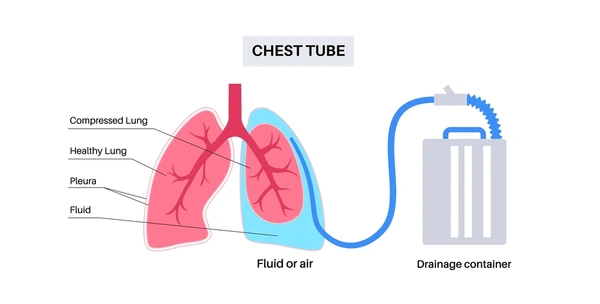
This procedure plays a vital role in managing pneumothorax, hemothorax, or pleural effusion by inserting a flexible tube for draining air, blood, or fluid. Continuous monitoring and meticulous care are imperative for ensuring effective drainage. Healthcare providers remain vigilant, promptly addressing complications to guarantee optimal patient recovery and preserve respiratory function.
Why Do I Need a Chest Tube?
Common reasons why a chest tube is needed include:
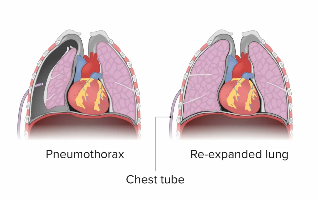
- Pneumothorax: A chest tube may be required to treat a pneumothorax, where air accumulates in the pleural space, causing a lung to collapse..
- Hemothorax: In cases of hemothorax, where there is blood accumulation in the pleural cavity, a chest tube helps drain the blood and restore normal lung function.
- Pleural Effusion: Chest tubes are often necessary for draining excess fluid in the pleural space, a condition known as pleural effusion.
- Post-Surgery Drainage:Following certain chest or cardiac surgeries, a chest tube may be inserted to facilitate drainage and prevent the accumulation of fluids in the pleural space.
Risks of Chest Tube Insertion:
- Infection: There is a risk of infection at the insertion site or within the pleural space, requiring careful sterile techniques during the procedure.
- Bleeding: Chest tube insertion carries the potential for bleeding, particularly if there is an injury to blood vessels during the procedure..
- Pain and Discomfort:Patients may experience pain or discomfort at the insertion site or in the chest area following the procedure.
- Vasovagal Reaction: Some individuals may experience a vasovagal reaction, resulting in dizziness, fainting, or a drop in blood pressure during or after the procedure.
Will there be any pain or possible complications when the chest tube is removed?
When the doctor determines that you no longer need the chest tube, it will be removed. Usually, it can be taken out right at your bedside. There rarely is a need for sedation medication. You will be told how to breathe as the tube is being pulled. A secure bandage will be put in place. You will be told when the bandage can be removed. Often, a follow-up chest X-ray will be done to make sure that fluid or air hasn’t come back. Generally, there are no complications from the chest tube once it has been removed. You will only have a small scar. Chest tube insertion is a relatively safe procedure when done by Dr. Parthiv shah who is a pleural effusion specialist in Mumbai
What happens when the chest tube is in?
Most people will need to stay in the hospital the entire time the chest tube is in. You will be checked often for possible air leaks, plugging of the tube, and any breathing problems you may be having. Usually, you will be able to breathe more comfortably with the tube in place. Sometimes pain around the area where the tube enters the chest may cause you to take more shallow breaths. The nurse or doctor will tell you how much you can move around with the chest tube in place. Less and less fluid drainage in the collection device often means your lungs are improving. Sometimes the tube is clamped and left in place to make sure no fluid or air comes back before it is pulled out.
Chest tube insertion
Fluid or air in the chest that needs to be drained is identified using chest imaging such as chest X-ray, chest ultrasound, or chest CT scan. If the X-ray shows a need for a chest tube thoracostomy to drain fluid or air, the procedure is likely to be done by a surgeon, a pulmonary/critical care physician, or an interventional radiologist.
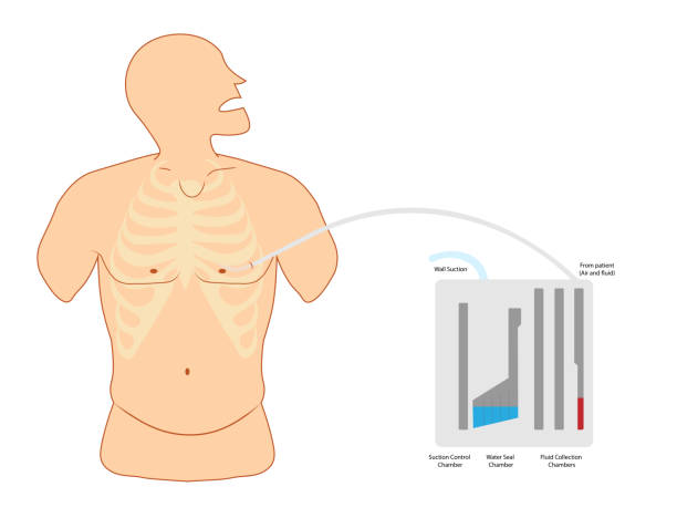
Often an adult or older child remains awake when a chest tube is inserted, except when it in place in the operating room during an open chest procedure. Sometimes a person, particularly a younger child, is given a small amount of medicine (a sedative) that causes sleepiness before a chest tube is inserted. The skin will be thoroughly cleaned. A local anesthetic (numbing) medication will be injected into the skin and in the tissue along the path between the ribs that the tube will follow. A cut (incision) from ¾ inch to 1½ inches long, between the ribs (the exact location depends on what is being drained and its location in the lungs). The chest tube is inserted and will be stitched into place to prevent it from slipping out. An airtight sterile dressing bandage is placed over the insertion site. The chest tube will be connected to a drainage collection device (usually a clear plastic container that rests on the floor). Often it is attached to suction to help draw out the air or fluid. Get treatment for pleural effusion by Dr Parthiv Shah- Pleural effusion specialist in mumbai.


 Alka Johari2023-12-24Dr More gives enough time to listen to the patient. His diagnosis is up to the mark and he very nicely explains the underlying health problem.
 pratibha bhanej2023-11-30Dr.More chest specialist at Dhanori is a very well known Doctor in Pune.His diagnosis and medications is very effective and accurate.Very few doctors say that I want my patience to be on least medicine and he avoids giving too many medicine. The clinic is clean and very hygienic.All tests done are reasonable and accurate. Suyog is also very helpful who does the test.Very systematic practice of giving appointments with reasonable fees.
 Kushal Sachdeva2023-11-10A Breath of Relief: Dr. Vishal Ramesh Moore, the Unmatched Master in Chest Illness I recently had the privilege of being under the care of Dr. Vishal Ramesh Moore, and I can confidently say that he is a true master in the realm of chest illness. My journey with a persistent and unstoppable cough led me to seek out a specialist who not only possessed exceptional medical expertise but also genuine compassion for his patients. Dr. Moore exceeded my expectations on both fronts. From the moment I stepped into his office, I was met with a warm and welcoming atmosphere. Dr. Moore's approachability and affable demeanor immediately put me at ease, fostering an environment where open communication was encouraged. Unlike some doctors who seem more focused on financial gain than patient well-being, Dr. Moore's primary concern was undoubtedly my health. What sets Dr. Moore apart is his ability to diagnose illnesses swiftly and accurately. In my case, he wasted no time in conducting a thorough examination and ordering relevant tests. His commitment to unraveling the root cause of my condition was evident, and I felt reassured knowing that I was in the hands of a dedicated professional. One of the most commendable aspects of Dr. Moore's practice is his genuine love for his profession. It's evident that he views medicine not merely as a job but as a calling. This passion translates into a level of care that goes beyond the standard, leaving patients like me grateful for having found such a dedicated and empathetic doctor. While my treatment is still ongoing, I am confident that it is heading in the right direction. Dr. Moore has been meticulous in tailoring a treatment plan specific to my needs, and the progress I've experienced so far is a testament to his expertise. Dr. Vishal Ramesh Moore is more than just a doctor; he's a beacon of hope for those grappling with chest illnesses. If you're in search of a healthcare professional who values your well-being over monetary gains and approaches their work with a passion for healing, Dr. Moore is the ideal choice. I am genuinely grateful to have found him and would wholeheartedly recommend his services to anyone in need of an exceptional chest illness specialist.
 Amol Agarwal2023-10-22I know Dr Vishal from 4+ years now. His diagnose is perfect. He is very down to earth person & is always approachable. Thanks for helping everytime doctor 🙏
 Shyam Gupta2023-10-21Dr. Vishal More, I would say excellent in treating chest illness, I went for cough treatment he done necessary testing and given required medicines. Much relief now. I must recommend Dr Vishal More because of my personal experience. Thank you Doctor.
 Saroj Kumar Nayak2023-09-05I have been consulting for my child since last 5 months. His night sleep was horrifying due to wheezing episodes. I remember on my first visit, Dr. waited beyond his timings to get the diagnosis done. Also I am quite fortunate to get instant response in initial days. It was great relief and much needed support. Now my child's health has improved and I am very optimistic with the medication process. Every time, he patiently listens to us and gives a lot of confidence to deal with the disease. His explanation and treatment for the disease is excellent. Also the clinic is well organised. Thank you Doctor.🙏 I would recommend Dr. Vishal More for anyone having respiratory problems.
 Abhimanyu Bhagat2023-08-29I have known Dr. More since past 4 years and he is a excellent patient advisor and consultant. He thoroughly listens to the patients views and his clinical diagnosis is very sharp. He also explains the condition and treatment patterns in a detailed and well manner which itself provides great relief. Also, the staff at the clinic is very friendly and professional with well organised appointment system. I’ve never had to wait more than a few minutes when I arrive on time for an appointment. I always felt confident and in safe hands in my receiving expert medical care. Whenever necessary, he also responds over the phone and ensures the right advice and treatment indicating his highly dedicated patient centric approach. I highly recommend him to anyone looking for a specialist. God bless him and best wishes for the future.
 Aroon Idnani2023-08-22I highly recommend Dr. Vishal More’s Clinic for their exceptional medical expertise and compassionate care. My experience with Dr. Vishal More is truly remarkable, as he took the time to listen to my issues and thoroughly explain the situation and provide excellent treatment options. The professionalism and dedication to patient well-being are truly commendable. I trust Dr. Vishal More wholeheartedly and believe he is an excellent choice for anyone seeking top-notch medical care regarding their chest and breathing related issues.
 Rutu Gandhi2023-08-22Very nice and supportive attitude
 Shruti Pillay2023-06-08Since childhood being around Doctors I had lost my trust in them overtime as I was sure that Doctors are just to loot. Dr. More helped me change that view, he is experienced and quick in identifying problems with the body, he treated my diabetic mother with utmost care for Tuberculosis. Kudos to Doctors like Dr. More.
